Article Text

Abstract
One in five strokes affects the posterior circulation. Diagnosing posterior circulation stroke can be challenging, as the vascular anatomy can be variable, and because presenting symptoms are often non-specific and fluctuating. Nevertheless, making the correct diagnosis is important, as these strokes have a high chance of recurrence, can be life threatening, and can lead to equally life-threatening complications. Investigation and management largely follow those for stroke in general, although some specific differences exist. These include the preferred use of MRI for diagnosing posterior fossa lesions, the management of basilar artery thrombosis, which may have a longer time window for recanalisation therapy, and the use of endovascular therapies for secondary prevention, which, so far, have not shown any benefit in the treatment of vertebral or basilar artery stenosis. In this review, we summarise the anatomy, aetiology and presentation of posterior circulation stroke, and discuss current approaches to management.
- STROKE
- CEREBROVASCULAR DISEASE
Statistics from Altmetric.com
Introduction
About one in five strokes affect the posterior circulation.1 Despite this relatively high incidence, posterior circulation stroke has received less attention and has often been managed differently compared to anterior circulation stroke. Potential causes include diagnostic difficulties, lack of non-invasive investigations and perceived differences in pathophysiology and risk. Diagnosing posterior circulation stroke can be challenging, as it often presents with non-specific symptoms, or symptoms that overlap with anterior circulation stroke or stroke mimics. Imaging of the posterior fossa and of the vertebrobasilar arterial system was difficult prior to MRI and non-invasive vascular imaging with CT or MR angiography (CTA, MRA) becoming more widely available. However, with the advent of modern imaging, it has been possible to study posterior circulation stroke in more detail, and interest in this field has grown.2–4 While often similar to the anterior circulation, there are certain aspects of the clinical presentation, potential complications and management of posterior circulation stroke that are important to be aware of for appropriate patient management. These were highlighted in recent reviews.3 ,4 Our current review provides an update, specifically addressing recent trial data for interventional treatment in vertebral artery stenosis, and we also focus in detail on presentation and management of basilar artery occlusion (BAO), and more unusual presentations of posterior circulation events.
Anatomy of the posterior circulation and important variants
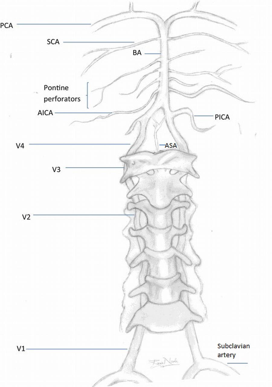
The posterior circulation is made up of the vertebral arteries, basilar artery, posterior cerebral arteries and their branches (figure 1). The vertebral arteries arise from the subclavian arteries, although can occasionally originate directly from the aortic arch. They are typically divided into four segments. The first three segments run extracranially: V1—from the origin to the entry into the transverse foramen; V2—the course through the transverse foramina; V3—a loop from C2 around the atlas to the dura. The intracranial segment (V4) runs from the dura to the upper medulla or lower pons. It gives off the posterior inferior cerebellar artery (PICA), which supplies parts of the medulla oblongata and cerebellum. Furthermore, each V4 segment gives off one small branch that unites with its contralateral counterpart to form the anterior spinal artery. Both vertebral arteries unite at the pontomedullary junction to form the basilar artery. The basilar artery runs along the anterior pons. It gives off multiple paramedian and short circumferential branches to the pons, the anterior inferior cerebellar artery (AICA) and the superior cerebellar artery (SCA), before dividing into the two posterior cerebral arteries (PCAs) at the ponto-mesencephalic junction.

Posterior circulation vessels. The drawing shows the four segments (V1–V4) of the vertebral arteries from their origins at the subclavian arteries. They give off the posterior inferior cerebellar arteries (PICA) and anterior spinal artery (ASA), before joining intracranially to form the basilar artery (BA). The basilar artery gives off pontine perforating vessels as well as the anterior inferior cerebellar artery (AICA), and the superior cerebellar artery (SCA). It then divides into the posterior cerebral arteries (PCAs). Adapted from Nouh et al,48 copyright under Creative Commons License.
Anatomical variants
Anatomical variants are common in the posterior circulation. While usually asymptomatic, it is important to be aware of them, as they may contribute to stroke risk, and may be of relevance when determining stroke aetiology.
Vertebral artery hypoplasia (figure 2A) is present in a quarter of the general population, but appears to be more common in people with posterior circulation stroke.5 Stroke risk may be increased due to asymmetrical flow dynamics, leading to development of shear stress and of atheroma.

Examples for anatomical variants in the posterior circulation. (A) Hypoplastic vertebral artery (arrow); (B) fetal origin posterior cerebral artery on the right (bold arrow), absent posterior communicating artery on the left (interrupted arrow); (C) bilateral thalamic infarcts, consistent with the presence of an artery of Percheron; (D) right vertebral artery ending in the posterior inferior cerebellar artery (bold arrow), the patient also has a stenosis of the V4 segment on the left (interrupted arrow).
In 10% of people, the P1-segment of the PCA (between the PCA origin and the posterior communicating artery) is absent, and the PCA receives its entire blood supply from the internal carotid artery. This variant is known as a fetal origin PCA (figure 2B). In such cases, an infarct in the PCA territory may indicate that an ipsilateral carotid stenosis is symptomatic.6
The prevalence of a further variant, an artery of Percheron, is unknown, as these vessels are too small to be visible on conventional imaging. If present, perforating branches to both medial thalami arise unilaterally from one PCA. Occlusion of an artery of Percheron will lead to bilateral infarction of the paramedian thalami, with or without rostral midbrain involvement (figure 2C).7
Other anatomical variants include hypoplasia or aplasia of the intracranial segment of the vertebral artery, which terminates in the PICA, without contributing to the blood supply of the brainstem (figure 2D). Hypoplasia of the basilar artery is rare, and often associated with bilateral fetal origin PCAs. The prevalence of a persistent trigeminal artery is 0.1–0.6%. This artery originates from the internal carotid artery after its exit from the carotid canal, and anastomoses with the midbasilar artery. The part of the basilar artery caudal to the anastomosis is usually hypoplastic.8
Aetiology of posterior circulation ischaemia
Causes for posterior circulation ischaemia include embolism from the heart or large artery atherosclerotic disease, small vessel disease or vertebral artery dissection.9 Atheromatous lesions are commonly located at the origin of the vertebral arteries, in the intracranial vertebral arteries, either at the point where the artery penetrates the dura, or at its junction with the basilar artery, and in the proximal and middle basilar artery. Atheroma can be a cause of emboli or of haemodynamic compromise, if severe occlusive lesions are present. Common sites for embolic occlusions are the intracranial vertebral artery (ICVA) and PICA, where they cause medullary or cerebellar infarction, and the distal basilar artery or PCAs, where they cause midbrain and occipital infarcts.9 ,10
Recurrent ischaemic events affecting the brainstem have for a long time been labelled as ‘vertebrobasilar insufficiency’ and been attributed to haemodynamic compromise in the presence of severe vascular occlusive lesions. However, in the New England Medical Centre Posterior Circulation Registry (NEMC-PCR), which studied 407 consecutive patients with posterior circulation stroke, haemodynamically sensitive atheromatous lesions were rare.10 In the rare subclavian steal syndrome, the subclavian artery is stenosed proximal to the vertebral artery origin. If the blood supply to the arm is insufficient, blood can be recruited by flow reversal in the ipsilateral vertebral artery with associated steal from the vertebrobasilar circulation and potential development of brainstem symptoms. In Bowhunter syndrome, haemodynamic symptoms occur when a vertebral artery is compressed during head rotation, for example, by a bony spur, and the contralateral vertebral artery is hypoplastic or occluded, making collateral supply insufficient.11
Vertebral artery dissection is a common cause of posterior circulation stroke in the young. It can be caused by trauma, but is often spontaneous. Dissections are often located in the V2 or V3 segments of the vertebral artery. Infarcts are generally due to embolisation of thrombotic material that has developed at the site of the vessel wall tear, rather than haemodynamic compromise from vessel occlusion. In 10% of cases, a dissection can extend intracranially, and may then cause a subarachnoid haemorrhage.4
In vertebrobasilar dolichoectasia, the arteries are elongated, widened and tortuous. Posterior circulation ischaemia may develop secondary to thrombus formation in the presence of reduced flow, with subsequent embolisation, or local vessel occlusion. Other potential complications include ischaemia due to distortion of the orifices of arterial branches, symptoms related to compression of the brainstem and cranial nerves, and vascular rupture with a catastrophic outcome.12
Fabry disease is a rare, X linked lysosomal storage disorder due to deficiency of the enzyme α-galactosidase A.13 Globotriaosylceramide accumulates in various tissues, including vascular cells, with subsequent development of a vasculopathy. In a large registry of patients with Fabry disease, stroke occurred in 4.3% of women and 6.9% of men, and was ischaemic in 87%. Some studies have reported a higher prevalence of posterior circulation stroke in Fabry disease. Basilar dolichoectasia is present in 56% of men and 35% of women with Fabry disease, and may be a useful marker for neurovascular involvement. Treatment consists of replacing the missing enzyme. However, there is currently no convincing evidence that this reduces the risk of cerebrovascular complications.
Giant cell arteritis (GCA) causes ischaemic stroke in approximately 7% of patients, predominantly affecting the vertebrobasilar circulation.14 However, the prevalence of coexisting other vascular risk factors in the reported studies was high, so there may not be a direct causal link. Treatment follows the standard treatment for secondary stroke prevention, and steroids are used to treat the GCA.
Clinical presentation
General symptomatology
Posterior circulation strokes can present with a large variety of symptoms and signs due to the proximity of brainstem nuclei and large afferent and efferent tracts. While classical posterior circulation syndromes are only rarely found,15 there are a number of symptoms and signs that are suggestive of posterior circulation ischaemia, and important to recognise. In the 407 patients from the NEMC-PCR, the most frequent presenting symptoms were dizziness (47%), unilateral limb weakness (41%), dysarthria (31%), headache (28%) and nausea or vomiting (27%). The most frequent signs were unilateral limb weakness (38%), gait ataxia (31%), unilateral limb ataxia (30%), dysarthria (28%) and nystagmus (24%).16 Most of these are non-specific for posterior circulation ischaemia, especially if they occur in isolation. Nevertheless, patients with a posterior circulation stroke experience isolated brainstem symptoms (brainstem transient neurological attacks (TNAs)) preceding their stroke significantly more frequently than patients with anterior circulation events, suggesting that these indicate posterior circulation ischaemia.17 A high level of suspicion and familiarity with the presenting features of infarcts in the different posterior circulation territories are therefore required to inform further management. The remainder of this section summarises clinical presenting features.
Intracranial vertebral artery, posterior inferior cerebellar artery
Occlusion of these vessels causes infarction in the medulla oblongata, and in the PICA-supplied parts of the cerebellum. The most common syndrome related to occlusion of the ICVA is the lateral medullary syndrome (Wallenberg's syndrome), which occurred either alone or in combination with other infarcts in 72% of cases with ICVA occlusive disease in the NEMC-PCR.2 ,18 Similarly, approximately 50% of cases with isolated lateral medullary infarction are associated with ICVA-disease.19 The multiple presenting features of this syndrome are outlined in table 1. In medial medullary infarction, patients commonly develop weakness, and sometimes sensory loss, of the contralateral arm and leg. If the entire hemimedulla is infarcted, patients will have a Wallenberg's syndrome with additional contralateral hemiparesis and sensory loss. Rarely, dissection of the ICVA may lead to occlusion of the anterior spinal artery and spinal cord infarction.
Lateral medullary syndrome: clinical presentation and affected brainstem structures
Infarction of the PICA-supplied part of the cerebellum may occur with or without associated lateral medullary involvement, and it may affect the medial, lateral or entire PICA territory. The most prominent feature of medial cerebellar infarction, with infarction of the vermis, is severe vertigo.20 This may be the only symptom, and can be very difficult to distinguish from a peripheral vestibulopathy. The Head Impulse—Nystagmus—Skew Test (HINTS) is a useful bedside test to help make this distinction, with reportedly 100% sensitivity and 96% specificity to identify a central cause of vertigo21 (table 2).
Details of the Head Impulse–Nystagmus–Skew Test to distinguish between vertigo caused by peripheral or by central lesions.21
Truncal ataxia is a further prominent feature of PICA territory infarction, which may be missed if the patient is only examined in bed and not asked to stand or at least sit up. When walking, patients usually veer to the side of the lesion. In lateral cerebellar infarction, patients usually also develop ataxia, but vertigo is a less prominent feature. If the entire PICA territory is affected, patients frequently develop headache, neck pain and vomiting as additional symptoms.20 ,22 They need to be monitored closely, as they are at risk of developing malignant cerebellar infarction.
Basilar artery
The basilar artery can be divided into the lower third—origin to AICA; the middle third—AICA to the SCA; and the upper third—SCA to bifurcation into PCAs. Commonly, occlusion of the lower and mid-basilar artery is due to atheroma, and distal occlusion due to embolism. In atheromatous occlusion, prodromal transient ischaemic attacks, TNAs, or minor strokes occur in approximately 20–40% of patients, which, if recognised, offer an opportunity to prevent recurrent events by instituting secondary prevention.23 ,24
The clinical presentation of BAO is variable, depending on the location and aetiology of the occlusion, vascular anatomy, and presence of collateral circulation. The clinical features of occlusion of the basilar artery and of its major branches are outlined in table 3.
Basilar artery occlusion and branch infarction—anatomy and clinical features
Posterior cerebral arteries
The PCAs have two main territories of vascular supply: the deep PCA territory is supplied by the proximal PCA branches, and includes the paramedian midbrain and the medial and posterolateral thalamus. The superficial PCA territory includes the occipital lobes and variable parts of the medial temporal and parietal lobes.25 The clinical presentation of PCA-infarction is variable, depending on the location of the vascular occlusion. Visual field defects are the most common presenting feature. Neuropsychological deficits will occur with temporal and with parietal lobe involvement. They include memory deficits, and various disconnection syndromes affecting the visual and language pathways. Examples include:
Alexia without agraphia: left PCA infarction affecting the splenium of the corpus callosum, and leading to disconnection of right visual pathways from the left hemispheric language.
Gerstmann syndrome: angular gyrus infarction, leading to acalculia, agraphia, finger agnosia, right-left disorientation.
Prosopagnosia: right-sided PCA-infarction, inability to recognise faces.
A common symptom of PCA-infarction is headache, which may lead to diagnostic confusion with migraine, especially if visual symptoms are present.25
Mimics and chamaeleons of posterior circulation stroke
Several conditions present with brainstem symptoms and signs and may mimic posterior circulation stroke. While a history of sudden symptom onset suggests a vascular aetiology, and is diagnostically helpful if present, it may not always be readily available. Furthermore, brainstem ischaemia can be of stuttering onset in the presence of severe atheromatous disease, and mimic other conditions, making a clinical diagnosis difficult.24
In patients presenting with vertigo, a peripheral vestibulopathy has to be distinguished from a cerebellar or brainstem infarct. The HINTS test (table 2) is clinically helpful, although brain imaging will often be used for confirmation.
Central pontine myelinolysis (CPM) and Wernicke's encephalopathy present with brainstem deficits. A history of rapid correction of hyponatraemia, or of poor nutritional intake will clarify the diagnosis, but may not always be readily available. Basilar migraine is an important mimic of brainstem ischaemia, and may be distinguished by a more progressive symptom onset, and by a history of recurrent attacks. In posterior reversible encephalopathy syndrome (PRES) patients classically present with headache, seizures and encephalopathy. However, 10–15% of patients develop a focal neurological deficit, and due to involvement of the occipital lobes, visual defects are common and may mimic a posterior cerebral artery stroke. A background of severe hypertension, immunosuppression, renal failure or eclampsia, and imaging appearances of bilateral posterior subcortical vasogenic oedema, will help to clarify the diagnosis.26
In some cases, the clinical presentation of a posterior circulation stroke suggests a different diagnosis. Convulsive movements similar to seizures can be seen in brainstem and thalamic strokes, especially with pontine involvement. They can be severe, and a patient may mistakenly be diagnosed as having status epilepticus. The presence of pupillary and eye movement abnormalities should alert the clinician to the correct diagnosis.27
Investigation
The investigation of posterior circulation stroke and TIA follows this of stroke in general, including brain and vascular imaging, cardiac investigations, risk factor profiling and more specialised investigations as appropriate. MRI should be the preferred method for brain imaging, as CT imaging of the posterior fossa is prone to artefacts originating from the skull bone. In particular diffusion-weighted imaging (DWI) has a high sensitivity for acute ischaemic lesions, although can be falsely negative, especially for small posterior fossa infarcts.4 This was shown in a study of 356 patients, which compared CT and MRI in assessment for suspected acute stroke. Relative to the final diagnosis, the sensitivity and specificity of DWI were 83% and 96%, and of CT 16% and 97%. In this study, the odds were significantly higher for false negative DWI occurring in posterior versus anterior circulation events (OR=7.3; 95% CI 2.2 to 25.0).28 For vascular imaging, contrast-enhanced MR angiography (CE-MRA) is sensitive for diagnosing stenoses in the vertebrobasilar system. However, due to breathing artefacts, imaging of vertebral artery origin stenosis with CE-MRA can be difficult. With improving technology, CT angiography (CTA) is now probably aequivalent to CE-MRA, if not better, for imaging the posterior circulation.4 ,29 Further MRI and CT techniques can provide haemodynamic information and demonstrate collateral flow, for example, multiphase CTA. While currently mainly used in research settings, they may help patient selection for recanalisation treatment in the future. Duplex and Doppler ultrasound can be used to visualise the vertebral artery origin, and to demonstrate flow reversal in the vertebral arteries, and transcranial Doppler can show intracranial vascular occlusive lesions. However, Doppler is more operator dependent and less sensitive in the diagnosis of posterior circulation disease than MRA or CTA, and rarely used as the sole or primary investigation.4 ,10 ,29
Management
Most of the management of anterior and posterior circulation stroke and TIA is very similar, and outlined in recent guidelines.30–32 This section will cover the aspects of management that are specific to the posterior circulation.
Management of Basilar Artery Occlusion
Initial studies of BAO relied on autopsy findings, creating a perception that outcome was generally poor. This did not change with the advent of cerebral angiography, where mortality rates of 85% were still reported. Progress in non-invasive imaging has shown that BAO does not invariably lead to severe deficits. The international BASICS registry studied 592 patients with BAO. Of these, 347 (59%) presented with a severe deficit, defined as patients in a coma, with tetraplegia, or in a locked-in state, and 245 patients (41%) presented with a mild to moderate deficit, defined as any presentation other than severe. At 1 month, mortality in this group was 17%, and 38% were functionally independent, compared to 50% and 11%, respectively, in patients with a severe deficit at onset.33
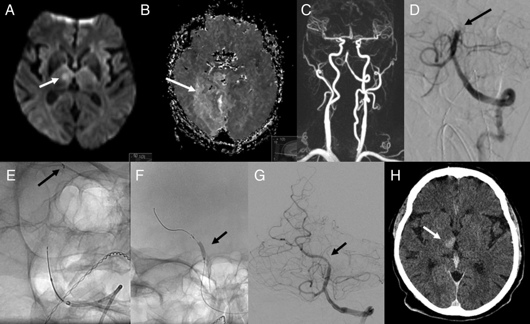
Outcome in acute BAO improves if recanalisation is achieved: In a meta-analysis of observational studies, recanalisation was associated with a relative risk of 0.67 (0.63 to 0.72) of death or dependency, and of 0.49 (0.44 to 0.55) of death compared to no recanalisation.34 However, while recanalisation rates were higher in the intra-arterial (77%; n=1715) compared to intravenous group (59%; n=341), outcomes between these two groups did not differ significantly. This may be explained by many studies in this review using first-generation retrieval devices, which also failed to show benefit compared to intravenous thrombolysis in anterior circulation stroke. Stent retrievers, which were used in the recent positive thrombectomy trials in anterior circulation stroke, were also found to be safe and associated with high recanalisation rates in acute BAO in a recent systematic case series review.35 Randomised data are still lacking, but the ongoing BASICS trial will compare the safety and efficacy of additional intra-arterial treatment in patients with persistent BAO after intravenous thrombolysis.36 An example of intra-arterial BAO recanalisation is shown in figure 3.

Basilar artery occlusion. Hyperacute ischaemic changes in the right thalamus on diffusion-weighted imaging (DWI) (A, arrow). There are no DWI changes affecting the right occipital lobe (A), but there is an extensive deficit on perfusion-weighted imaging (B, arrow). MR angiography (C) and intra-arterial angiography (D) confirm an occlusion of the mid-basilar artery, as well as an occluded right vertebral artery. The patient is treated with thrombolysis, clot retrieval (arrow indicates position of retrieval device), (E) and subsequent stent placement and balloon angioplasty of a severe underlying residual stenosis of the basilar artery at the occlusion site. (F) Recanalisation and re-establishment of flow in the basilar and right posterior cerebral arteries is achieved. There is a left fetal posterior cerebral artery, which does not fill from the basilar injection. (G) CT of the brain with contrast the following day (H) confirms the presence of a right thalamic infarct (arrow), but shows no additional ischaemic changes in the right occipital lobe, which was previously affected by the perfusion deficit (Images provided by Dr Pasquale Mordasini, Neuroradiology, Bern).
Several observational studies have used longer time windows for recanalisation therapy in BAO than in the anterior circulation. While time to treatment was a significant prognostic variable in the univariate analyses, it no longer was after adjusting for the extent of baseline ischaemic changes. A potentially longer time window for recanalisation in BAO could be explained by a better collateral supply in the posterior circulation, a persisting layer of plasma flow surrounding the clot, and reverse filling of the distal basilar artery. Rather than time to treatment, more accurate outcome predictors in BAO appear to be the achieving of recanalisation, severity of initial neurological deficit, extent of infarction on initial brain imaging, and presence of collateral supply.37
Malignant cerebellar infarction
In extensive cerebellar infarction, mass effect with compression of the brainstem and the fourth ventricle, subsequent development of hydrocephalus and brainstem herniation can occur. Studies suggest that approximately 20% of patients with cerebellar infarction develop some mass effect and associated neurological deterioration.22 While guidelines for management exist, these are based on a small number of case series. Data from randomised trials are lacking.22
The most reliable indicator for the development of tissue swelling is level of consciousness, although methods to monitor this accurately are still missing. Pontine compression can also cause eye movement abnormalities, respiratory depression and cardiac dysrhythmias. Swelling can take several days to develop. Imaging markers of malignant cerebellar infarction include effacement of the fourth ventricle as the key radiological indicator, followed by compression of the basal cisterns, brainstem deformity, hydrocephalus, downward tonsillar herniation, and upward transtentorial herniation. Generally, neurological deterioration is initially due to brainstem compression, with development of hydrocephalus a secondary effect. Medical management includes close observation, preferably in an intensive or high care environment. Intubation and ventilation may be required. Extreme hypertension should be avoided, but clear blood pressure targets have not been determined. Temporary reduction of tissue swelling with hyperosmotic agents may be reasonable, but steroids are not beneficial.22 Surgical posterior fossa decompression for malignant cerebellar infarction has been described as beneficial in case series, but prospective randomised data do not exist. The recommended surgical intervention is decompressive posterior fossa craniectomy, with or without extraventricular drainage and removal of infarcted tissue. Ventricular drainage alone is not recommended, due to the risk of upward herniation of the swollen cerebellum and brainstem. The time interval to surgery does not seem to affect outcome.22
Risk of recurrence and secondary prevention
The risk of recurrent ischaemic events after posterior circulation TIA or minor stroke is at least as high as in the anterior circulation.38 This may partly be explained by a high prevalence of atheromatous vascular stenosis, which may be higher in the posterior than in the anterior circulation.39 As in the anterior circulation, the risk of recurrence is high in the presence of stenotic disease. In vertebral artery stenosis, this risk differs according to plaque location. In a pooled analysis of 323 patients, the 90-day risk of recurrent stroke was 2.8% in patients with no stenosis, 5.4% in patients with extracranial vertebral artery stenosis (not significantly higher compared to patients without stenosis), and 13.9% in patients with intracranial stenosis (OR=5.6; 95% CI 1.7 to 18.7, p<0.0001).39
Medical treatment in secondary stroke prevention is the same for anterior and posterior circulation events and detailed in recent guidelines.31 ,32 It generally includes an antiplatelet agent, or anticoagulants in preceding cardioembolic events, cholesterol lowering drugs, and antihypertensive medication. However, in patients with severe vascular occlusive disease, who may have haemodynamic events, any blood pressure lowering should be done very cautiously. Subgroup analyses from trials of carotid endarterectomy indicate that in the anterior circulation, blood pressure lowering in patients with severe bilateral carotid disease increases the risk of stroke. It would be reasonable to assume a similar association in the posterior circulation, although no aequivalent studies exist.4
In symptomatic carotid stenosis, early endarterectomy or stenting reduces the risk of recurrent events. Surgical options to treat vertebral artery stenosis exist, but are rarely used. Endovascular approaches have been used more frequently over recent years, but until recently randomised data were lacking. Meta-analyses of non-randomised case series had found a low periprocedural risk of stroke (1.3%) or death (0.3%) with stenting of vertebral artery origin stenosis.40 ,41 In comparison, angioplasty or stenting for ICVA stenosis had higher risks of periprocedural stroke (10.3% after stenting, 7.6% after angioplasty) and death (3.2% and 3.7% respectively).40
In CAVATAS, only 16 patients with vertebral artery stenosis were randomised either to medical treatment alone (N=8) or angioplasty (N=8). All patients had extracranial stenosis, and no patient had an outcome event during 4.7 years of follow-up. Recruitment late after the presenting event and small patient numbers do not allow firm conclusions to be made.42 The Vertebral Artery Stenting Trial (VAST), an open-label randomised trial of stenting plus best medical treatment in recently (<6 months) symptomatic vertebral artery stenosis versus best medical treatment alone was stopped early because of new regulatory requirements. In total 57 patients were assigned to stenting and 58 to medical treatment alone. Three patients in the stenting group had vascular death, myocardial infarction, or any stroke within 30 days of treatment (5%, 95% CI 0% to 11%) versus one patient in the medical treatment group (2%, 0% to 5%). During a median follow-up of 3 years, seven (12%, 95% CI 6% to 24%) patients in the stenting group and four (7%, 2% to 17%) in the medical treatment group had a stroke in the territory of the symptomatic vertebral artery. While outcome numbers were small, the authors concluded that the low risk of recurrent events on medical treatment alone questioned the need for and feasibility of a larger phase 3 trial.43 The Vertebral Artery Ischaemia Stenting Trial (VIST) also compares intervention with best medical treatment alone in recently (<3 months) symptomatic vertebral artery stenosis. Funding was recently withdrawn due to slow recruitment, but follow-up continues.44 The SAMMPRIS trial compared stenting with the Wingspan Stent versus aggressive medical treatment alone in patients with symptomatic intracranial stenosis. Medical treatment consisted of aspirin 325 mg daily indefinitely, clopidogrel 75 mg daily for 90 days, tight blood pressure control (<140 mm Hg systolic, or <130 mm Hg in diabetes), cholesterol lowering, aiming for LDL cholesterol <70 mg/dL (1.8 mmol/L), and a lifestyle modification programme. The trial was stopped early after recruiting 451 patients, due to a higher 30-day risk of stroke or death in the stenting group (14.7%) compared to the medical group (5.8%, p=0.002).45 A recently published subgroup analysis confirmed that the better 2-year outcomes with medical treatment did not differ significantly between anterior and posterior circulation, or across individual blood vessels. In SAMMPRIS, the 2-year risk of stroke or death was 9.5% (2.5% to 33.0%) for 22 patients with intracranial vertebral artery stenosis in the medical treatment arm, compared to 21.1% (11.1% to 37.7%) for 38 patients who underwent stenting. There were 51 patients with basilar artery stenosis receiving medical treatment alone, and 49 patients who underwent stenting. Their 2-year risk of stroke or death was 9.9% (4.8% to 19.4%) and 24.5% (14.7% to 39.1%), respectively.46 In the VISSIT trial, treatment with a balloon-expandable stent was compared to best medical treatment for symptomatic intracranial artery stenosis. Outcomes were reviewed after the results from SAMMPRIS had become known, and the trial was stopped early due to futility after recruiting 112 of 250 planned patients. The 30-day primary safety end point occurred more frequently in the stenting group (24.1%) versus the medical group (9.4%, p=0.05), as did the 1 year primary outcome of stroke or hard TIA (36.2% vs 15.1%; p=0.02). Outcomes specifically for posterior circulation stenosis are not available.47
The lack of benefit of intervention in these trials may partly be due to late recruitment in CAVATAS and VAST, at which point in time the high risk of early recurrence had already passed. However, the trials also showed a lower than expected risk on medical treatment, and the benefit of medical treatment over stenting persisted beyond the periprocedural period. The currently available data suggest that endovascular treatment of posterior circulation stenosis confers no benefit over medical management.
Future research
The best strategies for acute management and secondary prevention in posterior circulation stroke remain to be determined. The BASICS registry challenged the view that intra-arterial treatment conveyed additional benefit over intravenous thrombolysis in BAO,33 and randomised evidence is still lacking. Given the marked benefit of thrombectomy in the anterior circulation, patient recruitment for trials of thrombectomy in BAO may be challenging, but one such trial is ongoing.36 Further studies should address whether the time window for intervention differs in brainstem strokes and BAO compared with the anterior circulation. Future studies of intervention in vertebrobasilar stenotic disease may clarify differences in benefit between patients with haemodynamic compared to embolic events. New imaging strategies, for example, non-invasive imaging of collateral flow, need to be assessed in their usefulness to identify patients likely to benefit from acute or preventive interventional procedures, and target treatments appropriately.
Conclusion
The anatomy of the posterior circulation is variable, and presenting symptoms of ischaemic events are often non-specific and fluctuating. Diagnosing posterior circulation strokes can therefore be challenging, but is important, as these strokes have a high chance of recurrence, and can be life threatening. Investigation and management largely follow those for stroke in general, although specific differences exist. These include the preferred use of MRI for diagnosing posterior fossa lesions, the management of BAO, which may have a longer time window for recanalisation therapy, and the use of endovascular therapies for secondary prevention, which, so far, have not shown any benefit in the treatment of vertebral artery stenosis. Future studies should address the usefulness of intervention in acute and preventive treatment of posterior circulation stroke, and develop imaging strategies to help target treatments appropriately.
Acknowledgments
The authors would like to thank Dr Pasquale Mordasini, Institute of Diagnostic and Interventional Neuroradiology, Inselspital Bern, Switzerland, for contributing the images for figure 3.
References
Footnotes
Contributors US reviewed the literature and drafted the manuscript. UGF reviewed and commented on the manuscript and co-wrote the sections on diagnosis and management of basilar artery occlusion.
Funding US is part funded by the NIHR Biomedical Research Centre.
Competing interests None declared.
Provenance and peer review Commissioned; externally peer reviewed.