Article Text

Abstract
Background Haemorrhages of brainstem cavernous malformations (CMs) can lead to neurological deficits, the natural history of which is uncertain. The study aimed to evaluate the neurological outcomes of untreated brainstem CMs and to identify the adverse factors associated with worsened outcomes.
Methods From 2009 to 2015, 698 patients (321 women) with brainstem CMs were entered into the prospective cohort after excluding patients lost to follow-up (n=43). All patients were registered, clinical data were collected and scheduled follow-up was performed.
Results After a median follow-up of 60.9 months, prospective haemorrhages occurred in 167 patients (23.9%). The mean modified Rankin Scale scores at enrolment and at censoring time were 1.6 and 1.2. Neurological status was improved, unchanged and worsened in 334 (47.9%), 293 (42.0%) and 71 (10.2%) patients, respectively; 233 (33.4%) recovered to normal levels. Lesions crossing the axial midpoint (relative risk (RR) 2.325, p=0.003) and developmental venous anomaly (DVA) (RR 1.776, p=0.036) were independently significantly related to worsened outcomes. The percentage of worsened outcomes was 5.3% (18 of 337) in low-risk patients (neither DVA nor crossing the axial point) and increased to 26.0% (13 of 50) in high-risk patients (with both DVA and crossing the axial point). The percentage of worsened outcomes significantly increased as the number of prospective haemorrhages increased (from 1.5% (8 of 531, if 0 prospective ictus) to 37.5% (48 of 128, if 1 ictus) and 38.5% (15 of 39, if >1 ictus)).
Conclusions The neurological outcomes of untreated brainstem CMs were improved/unchanged in majority of patients (89.8%) with a fatality rate of 1.7% in our cohort, which seemed to be favourable. Radiological features significantly predicted worsened outcomes. Our results provide evidence for clinical consultation and individualised treatment. The referral bias of our cohort was underlined.
- vascular malformation
- brain
- haemorrhage
This is an open access article distributed in accordance with the Creative Commons Attribution Non Commercial (CC BY-NC 4.0) license, which permits others to distribute, remix, adapt, build upon this work non-commercially, and license their derivative works on different terms, provided the original work is properly cited, appropriate credit is given, any changes made indicated, and the use is non-commercial. See: http://creativecommons.org/licenses/by-nc/4.0/.
Statistics from Altmetric.com
Introduction
Cerebral cavernous malformations (CCMs) account for 10%–15% of intracranial vascular malformations, with a population incidence ranging from 0.4% to 0.8%.1 Due to their sluggish blood flow, they are often considered clinically quiescent and angiographically occult, and up to 40% of patients are asymptomatic. In a population-based cohort,2 the 5-year risk of a composite outcome (haemorrhage or focal neurological deficit (FND)) was approximately 57.1% for brainstem cavernous malformations (CMs), which was in accordance with the rate of 50.7% with haemorrhage/FND presentation in our recent meta-analysis.3 Due to the presence of vital and dense neurostructures, the eloquent site with a small volume and the high susceptibility to minor haemorrhage,4 the brainstem has been confirmed to be associated with symptomatic haemorrhage.3 5–7 The natural history of CCMs and favourable surgical outcomes of brainstem CMs have been extensively delineated,2 3 5 6 8–21 but there is a paucity of data regarding the neurological outcome of untreated brainstem CM, which has otherwise been poorly individually reported. Haemorrhage of brainstem CMs can result in severe neuropathies, impairments in quality of life and even unexpected death relevant to patients. Therefore, we aimed to describe the neurological status of untreated brainstem CMs in a prospective cohort and to review the literature regarding the untreated disease in order to contribute to improving our understanding of the natural history of the disease, to distinguish between patients who might suffer from worsened outcomes and subsequently become surgical candidates, and to facilitate clinical decision-making.
Patients and methods
Patient population
Patients diagnosed with brainstem CMs in our institute from January 2009 to December 2015 were registered and enrolled in the consecutive prospective cohort according to the inclusion and exclusion criteria. The inclusion criteria were (1) a definitive diagnosis of an untreated brainstem CM and (2) provision of informed consent. Patients were excluded if they met one of the following criteria: (1) the diagnosis was questionable/undefined; (2) they did not consent to follow-up; (3) they received treatment (radiation or surgery) before enrolment; (4) they had severe comorbidities involving the eloquent location, which might potentially influence the accuracy of neurological evaluation; (5) they were pregnant or breast feeding; and (6) they underwent surgery immediately following diagnosis. Surgical indications have been described in prior studies,15–19 22 23 and stereotactic radiosurgery (SRS) was not recommended. Surgical treatment was recommended if as follows: (1) multiple haemorrhages and poor or progressively aggravated neurological status; (2) lesion size ≥2 cm or enlarged with a significant mass effect; and (3) lesion grew exophytically or became abutting the pial surface after haemorrhagic ictus. All clinical data were obtained from a prospective clinical trial (no. ChiCTR-POC-17011575). Written informed consent was obtained from all participants and their parents or caregivers.
Procedures and neurological evaluation
The inception point of the study was the date of the first medical consultation or clinical visit that led to the diagnosis of a brainstem CM at our institute. Two neuroradiologists independently reviewed the MRI scans to verify the diagnosis according to the accepted criteria (online supplemental figure 1).13 In patients with MRI scans obtained more than 2 weeks prior, MRI re-examination was prescribed at enrolment to check the radiological features because haematomas evolve over time. The characteristics of MRI, including (but not limited to) an equivalent lesion size, crossing the axial midpoint,16 the Zabramski classification13 and developmental venous anomalies (DVAs), were extensively reviewed. If there were multiple CMs involving the brainstem, the symptomatic haemorrhagic CM took precedence for analysis; otherwise, the larger CM was preferred since both were postulated to be predictors of haemorrhage or FND.2 Brainstem CMs often encompassed different compartments, and we classified each CM only to one compartment according to the location of the maximal axial cross-sectional area of the lesion regardless of the number of anatomical compartments involved by the lesion (online supplemental figure 2). If the maximal axial cross-sectional area of a CM was located in the thalamus, the lesion would be classified as a thalamic CM even if it concurrently encompassed the midbrain. Neither the anatomical compartment within the brainstem nor the number of compartments involved by a brainstem CM had been proven to be related to the worsened outcome that had been observed in prior studies.15 16 18–22 Our classification was easily understandable, practicable and repeatable in clinical practice that conformed to the gross anatomy of the brainstem and facilitated statistical analysis.
Supplemental material
The initial presentation, number of prior haemorrhages, and symptoms and neurological status following each ictus were collected and identified according to the medical records combined with available radiographic evidence that facilitated judgement of the presence of haemorrhagic events. Cranial nerve palsy, long tract deficits (motor and sensory abnormalities) and/or extrapyramidal symptoms specifically pertinent to the anatomical location of the lesion were classified as FNDs. Headache, dizziness, nausea, vomiting and/or other symptoms not directly associated with the lesion were classified as non-FNDs (non-specific symptoms). The definition of haemorrhage according to that advocated by Al-Shahi Salman et al24 is as follows: a clinically symptomatic event (an acute or subacute, new-onset or worsening FND corresponding to the anatomical location of the brainstem CM, impaired consciousness, or severe headache) with recent overt haemorrhage within or outside the lesion leading to changes in lesion size and/or signal intensity based on serial radiographs.2 22 The first 3 days since onset of a clinically symptomatic event was defined as acute, and after that the interval ranging from the fourth day to 3 weeks was defined as subacute. Symptomatic events, provided by patients’ impression, recall or only medical records but without concomitant radiographs, were not enough to conclude a haemorrhagic event, which was considered uncertain to reduce recall bias and to avoid overestimating prior haemorrhages. Acute-onset symptoms or worsened deficits, anatomically consistent with the CM location, likely raised the suspicion of a haemorrhage, but other causes (ischaemic attack, infarction or secondary perilesional oedema) were not precluded in the absence of timely radiographs.
Follow-up
At enrolment, at least three phone numbers from the patients and their relatives were registered, along with information on home address, email address and social media account (Tencent Messenger and WeChat) if available. Two authors (PPL and ZYW), blinded to the results of the radiographs and haemorrhagic status, independently evaluated the neurological status at enrolment using modified Rankin Scale (mRS) scores and then completed the scheduled follow-up by clinical revisits (in person) 3, 6 and 12 months after enrolment and then annually after that or by telephone interviews, with visual contact according to structured question approaches or mailed questionnaires with digital photos sufficient for clinical evaluation. Home visits or connections with family doctors from community health services or community hospitals were alternative approaches to obtain follow-up information, with any discrepancy resolved by the senior author (JTZ). Since 2009, with the economic development, few patients would change their phone numbers, and these three phone numbers were less likely to change at the same time. These phone numbers and information for contacting the patients would be updated at each scheduled clinical revisit, and if clinical revisits were unavailable telephone interviews were performed annually to check the availability of these contact methods. MRI was recommended at the same time, but MRI at any time was necessary once the patient perceived severe headache or new/worsened pre-existing symptoms. Provided changes in neurological function, we were fully aware of a new haemorrhage, but it would be noted as questionable without radiological evidence.
The follow-up duration began at enrolment and ended on the date of surgery, SRS, death or the most recent evaluation, whichever occurred first. The cause of death was enquired from their relatives if the patient has passed away.
Statistical analysis and literature review
The clinical and baseline data stratified by neurological outcomes were compared (table 1). The risk factors for worsened outcomes were evaluated by univariate and multivariate binary logistic regression (method: Enter). Statistical analyses were performed using SPSS statistical package software (V.25.0), with significance set at p<0.05. A systematic review of the literature was performed regarding neurological outcomes in untreated brainstem CMs. Studies were reviewed and screened by two independent review authors using the terms ‘cavernoma’, ‘cavernous malformation’ and ‘cavernous hemangioma’ in the following databases from January 1984 to December 2019: EMBASE, PubMed, Cochrane, Ovid/Medline, Web of Science and EBSCO. We further reviewed all of the references provided in the identified studies and incorporated all pertinent citations. Studies that did not provide neurological outcomes were excluded.
Baseline and clinical data of brainstem cavernous malformations stratified by neurological outcome
Results
Patient demographics
During the period from 2009 to 2015, after excluding 43 patients lost to follow-up, 698 patients (321 women, 46.0%) with brainstem CMs referred to our institute were entered into the consecutive prospective cohort, with a mean age of 38.3±13.8 years. Nine patients (1.3%) had a family history and 96 patients (13.8%) had a history of hypertension. The mean duration of symptoms before enrolment was 21.3±44.7 months, and 680 patients (97.4%) had a prior haemorrhage.
At enrolment, 508 patients (72.8%) presented with a recent haemorrhage (188 acute and 320 subacute haemorrhage), 589 patients (84.4%) complained of FNDs, including cranial nerve deficits (n=466, 66.8%), long tract impairments (n=428, 61.3%) and extrapyramidal symptoms (n=177, 25.4%), and 197 patients (28.2%) had non-specific symptoms. The mRS scores at enrolment were 0, 1, 2, 3, 4 and 5 in 47 (6.7%), 400 (57.3%), 104 (14.9%), 66 (9.5%), 68 (9.7%) and 13 (1.9%) patients, respectively. The mean and median mRS scores were 1.6 and 1.0. Overall, 447 patients (64.0%) were able to perform usual activities, 104 (14.9%) had self-care abilities without the need for assistance, and 147 (21.1%) were unable to carry out self-care and required assistance. Radiologically, the pons (n=442, 63.3%) was the most common lesion location, followed by the midbrain (n=154, 22.1%) and medulla (n=102, 14.6%). The mean lesion size was 1.5±0.7 cm. The details of other radiographic parameters are described in table 1.
Follow-up
During a mean follow-up of 56.9±35.3 months (median 60.9 months), 167 patients (23.9%) suffered prospective haemorrhages. The censoring events (n=199, 28.5%) included SRS (n=14, 2.0%), surgical resection (n=170, 24.4%), disease-specific deaths (n=11, 1.6%) and deaths due to unrelated/unknown causes in the observation group (n=4, 0.6%).
Primary results: neurological status and risk factors for worsened outcomes
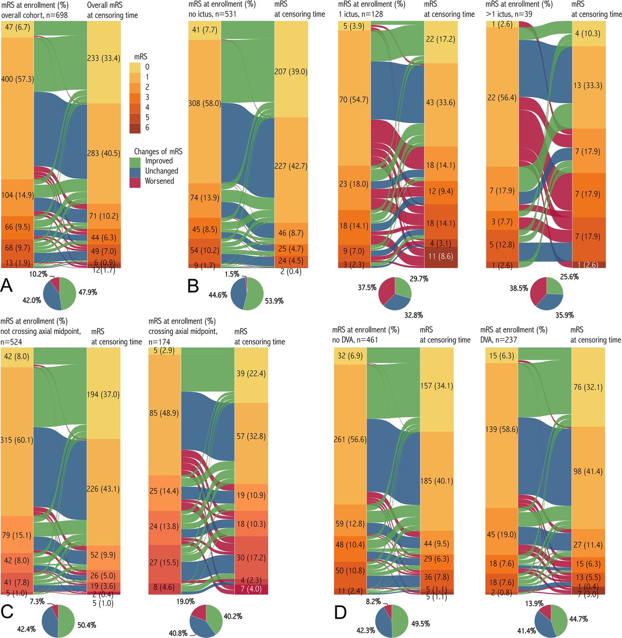
The mRS scores at censoring time were 0, 1, 2, 3, 4, 5 and 6 in 233 (33.4%), 283 (40.5%), 71 (10.2%), 44 (6.3%), 49 (7.0%), 6 (0.9%) and 12 (1.7%) patients, respectively. The mean and median mRS scores were 1.2 and 1.0. Compared with the mRS scores at enrolment, those in 334 patients (47.9%) improved, unchanged in 293 patients (42.0%) and worsened in 71 patients (10.2%) at censoring time (Z=−9.134, p<0.001) (table 1, figures 1 and 2). In 169 patients who were recommended surgery but at first refused, 52 (30.8%) suffered a prospective haemorrhage, 88 (52.1%) finally received surgery, and overall neurological status at censoring time was improved, unchanged and worsened in 59 (34.9%), 79 (46.7%) and 31 (18.3%) patients, respectively.

Sankey diagrams showing the distribution and changes in mRS score at enrolment and at censoring time. (A) The figure consisted of two bars and one flow in between. These two bars show the distribution of mRS scores at enrolment and at censoring time in orange. The width and value of each flow were calculated according to the sample size showing the dominant or subordinate contributions to the overall flow that were similar in other panels. (B–D) The mRS scores at enrolment and at censoring time were stratified by the number of prospective haemorrhages (B), crossing the axial midpoint or not (C), and presence of DVA or not (D). The Sankey diagrams are illustrated using Tableau Desktop (V.8.3). DVA, developmental venous anomaly; mRS, modified Rankin Scale.
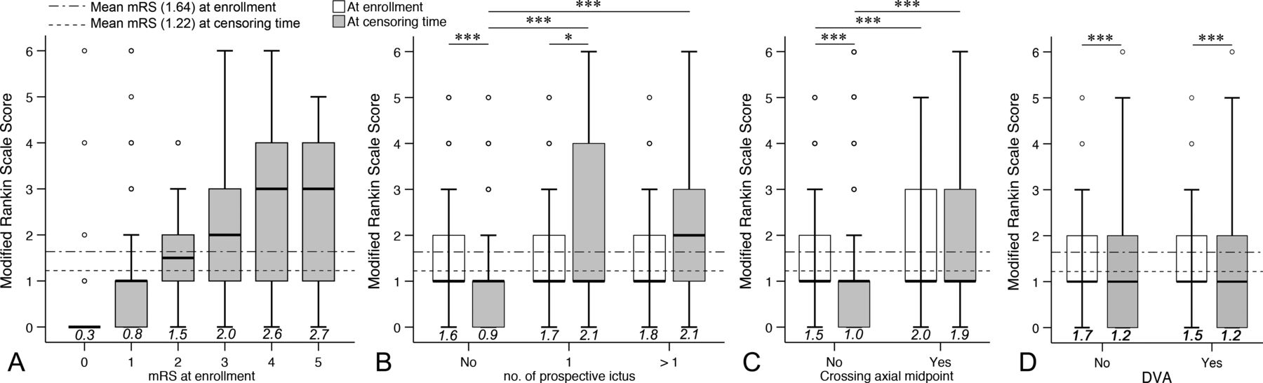
Boxplots showing the distribution of mRS scores at enrolment and at censoring time based on risk factors. (A) The boxplot shows the mRS score at censoring time stratified by mRS score at enrolment. (B–D) The outcome was significantly better in patients without prospective ictus than in patients with one (Z=−7.398, p<0.001) or more (Z=−5.414, p<0.001) ictus (B). Patients without lesions crossing the axial midpoint had significantly better outcomes than patients with lesions crossing the axial midpoint (Z=−6.352, p<0.001) (C). However, there was no significance between the outcomes in patients with or without DVA (Z=−0.384, p=0.701) (D). *P<0.05, ***P<0.001. Circles indicate the outliers; values in italic above the horizontal axis indicate the mean values of each boxplot. DVA, developmental venous anomaly; mRS, modified Rankin Scale.
The univariately significant risk factors for worsened outcome are lesion size (per 1 mm) (relative risk (RR) 2.343, p<0.001), crossing the axial point (RR 2.993, p<0.001), DVA (RR 1.801, p=0.020) and depth (RR 1.595, p=0.021) (table 2). In the multivariate analysis (Enter method) with adjustment for duration of follow-up, crossing the axial point (RR 2.325, 95% CI 1.332 to 4.060, p=0.003) and DVA (RR 1.776, 95% CI 1.037 to 3.041, p=0.036) were significantly related to worsened neurological outcomes (figures 1 and 2), and other factors were no longer significant. The percentage of worsened outcomes was 5.3% (18 of 337) in low-risk patients (neither DVA nor crossing the axial point), 12.9% (40 of 311) in mid-risk patients (either DVA or crossing the axial point) and 26.0% (13 of 50) in high-risk patients (with both DVA and crossing the axial point).
Risk factors for worsened neurological function in brainstem cavernous malformations
Secondary results: prospective haemorrhages and worsened outcomes
Overall, the numbers of patients experiencing 1, 2, 3 and 4 prospective haemorrhages were 128 (18.3%), 27 (3.9%), 8 (1.1%) and 4 (0.6%), respectively. The percentage of worsened outcomes significantly increased as the number of prospective haemorrhages increased (from 1.5% (8 of 531, if 0 ictus) to 37.5% (48 of 128, if 1 ictus) and 38.5% (15 of 39, if >1 ictus)), while the percentage of complete recovery significantly decreased from 39.0% (207 of 531, if 0 ictus) to 17.2% (22 of 128, if 1 ictus) and 10.3% (4 of 39, if >1 ictus) (χ2=31.965, p<0.001) (figures 1 and 2).
Discussion
The natural history of CMs has been elaborately demonstrated,1–3 5 6 8–13 but in prior studies brainstem CMs were often reported together with CMs of other locations rather than as a distinct entity, and the neurological outcomes of brainstem CMs were often not detailed or not described separately with limited sample size. Overall, the 5-year risk of a composite outcome (haemorrhage or FND) is 44.5% for brainstem CMs and 8.8% for CMs in other locations,3 the posthaemorrhage full recovery is 38.8%/person-years, and the haemorrhage-associated mortality is 2.2%.6 Due to the crucial anatomical location of the brainstem and unpredictable haemorrhage, the outcome of brainstem CMs is considerably poor. In our cohort, after a median follow-up of 5.1 years, we found that 233 patients (33.4%) recovered to an asymptomatic status, 334 (47.9%) improved and 71 (10.2%) worsened; additionally, DVA and crossing the axial midpoint had substantial effect on worsened outcomes. These results indicated that neurological status most likely trended towards improved/unchanged if without risk factors during the remaining lifetime of the patients.
Neurological outcomes of untreated brainstem CMs
Due to the relative rarity of brainstem CMs, they are often reported along with other locations with various outcomes. Given the aggressive features in surgical candidates, the preoperative complete recovery rate of 16.7% (6 of 36)25 was lower than that of observational series (25.9%–43.3%)22 23 26 and our cohort (33.4%). The complete recovery rate further declines following repeated haemorrhage.22 23 25 Across 16 prior studies and 1 meta-analysis with available data regarding the outcomes of untreated brainstem CMs (table 3),2 3 5 8 12 14 20–23 26–32 9 studies reported the evolution of neurological status compared with the initial status,12 20 22 23 27–29 31 32 6 studies only described the final status,2 5 14 21 26 30 and the remaining 2 studies did not describe the outcome.3 8 Several studies reported relatively poor outcomes, with a worsened proportion up to 41.7%,20 48.1%27 and 53.3%.28 Furthermore, Gross et al5 observed a permanent neurological morbidity rate of 45% for brainstem, thalamic and basal ganglia CMs; Al-Shahi Salman et al2 reported that 57.1% (8 of 14) of patients suffered a second composite outcome (haemorrhage or FND), and the worst outcome was underlined with an observational mortality rate of 20.0% (6 of 30).26 In contrast, three studies exclusively dedicated to the natural history of brainstem CMs reported fairly high percentages of improved outcomes, ranging from 38.8% to 59.8%12 22 23; our result (47.9%) was within this interval and was in accordance with that of a prior study (48.6%).12 In addition, Zimmerman et al,30 Kupersmith et al,12 Esposito et al32 and Bhardwaj et al31 reported favourable outcomes, with worsened percentages of only 12.5%, 8.1%, 5.9% and 7.7%, respectively. Overall, we performed a pooled analysis of nine studies,12 20 22 23 27–29 31 32 and the proportion of improved/unchanged and worsened outcomes was 85.2% (465 of 546) and 14.3% (78 of 546), respectively, which was analogous to our series and appeared more benign than previously thought.26–28 In 16 prior studies (n=685),2 5 8 12 14 20–23 26–32 disease-specific deaths occurred in 12 patients (1.8%),20 22 26 27 30 which was comparable with our series (1.7%, 12 of 698) and seemed to be acceptable. For censoring events, 24.4% of our patients underwent surgery, which was similar to a prior study (23.6%, 69 of 292)10 but higher than another cohort (17.2%, 23 of 134).2 The variation and wide spectrum of these outcomes might be attributed to the small sample size, selection and referral biases, different study designs, and diverse radiological parameters, which were also substantially vulnerable to prospective haemorrhages. Data on brainstem subsets extracted from prior studies were less convincing, but the results of pooled analysis would improve the level of evidence.
Neurological outcome of untreated brainstem CMs
Adverse factors for worsened/poor outcomes in brainstem CMs
The risk factors for outcomes were arguable and inconsistent in prior studies. In surgical series, larger lesion size,15 33 older age (eg, ≥12 years,17 >40 years19 per 5 years,16 and increased age),18 multiple haemorrhages,17–19 DVA16 and poor preoperative status17–19 were demonstrated to predict poor outcomes. However, opposing findings were occasionally reported in that multiple haemorrhages did not significantly lead to worsened outcomes.15 21 33 Parenchyma transgression, manipulation of circumferential tissue, poor postoperative neural plasticity and unfavourable baseline data might account for the variation.
Among untreated series, Kupersmith et al12 stated that the total number of episodes or rebleeding had little effect on neurological status. Conversely, Samii et al25 declared a lower chance of neurological recovery following multiple haemorrhages (6.25%) compared with only one episode (25.0%). A similar result reported by Li et al17 showed full recovery rates of 26.9%, 13.0% and 0% after 1, 2 and 3 haemorrhages, respectively. A new haemorrhagic episode would interrupt the course of recovery or would further lead to clinical deterioration. Our univariably significant risk factors were consistent with three of five predictors from the proposed grading scale (size, crossing the axial midpoint and DVA) advocated by Garcia et al.16 In our multivariable analysis, DVA and crossing the axial midpoint were independently significant. Lesions crossing the axial midpoint with mass effect, involving bilateral neurostructures and producing more morbidities, might restrain recovery after haemorrhagic ictus. DVA was found to be related to the genesis of CM and was also identified as a predictor of prospective haemorrhage; meanwhile, DVA might affect perilesional venous drainage followed by brainstem oedema, thrombosis or ischaemia, which jeopardised neurological function.5 34 Our results seemed to be applicable to clinical practice because both factors could be evaluated at enrolment.
Limitations of the study
Referral and selection biases were the main limitations of the study, which might overestimate the worsened outcomes and limit extrapolation of our findings to patients from other institutes. Patients with haemorrhagic CM with serious deficits were more likely referred for medical consultation due to survival instinct and the significantly unbalanced surgical techniques within our country. Despite the size of our cohort, this was not a true natural history because some patients already had a known diagnosis, while many others eventually underwent surgical treatment. Our findings were meaningful for patients who suffered from ruptured brainstem CMs but did not require immediate surgery or experienced hesitation regarding the selection of surgery. To minimise referral bias, a multicentre study or a population-based cohort is recommended based on the true natural history being theoretically more benign than in our cohort.
Conclusion
The neurological outcomes of untreated brainstem CMs were improved/unchanged in majority of patients (89.8%) and seemed to be favourable, and radiological features significantly predicted worsened outcomes. Our results provide updated evidence for clinical consultation and help to individualise patient treatment. Patients with risk factors require close follow-up. The referral bias in our study should be stressed.
References
Supplementary material
Supplementary Data
This web only file has been produced by the BMJ Publishing Group from an electronic file supplied by the author(s) and has not been edited for content.
Footnotes
DL, J-JZ and J-CW contributed equally.
LW and ZW contributed equally.
Contributors Conception and design: all authors. Acquisition of data: DL, Z-YW, P-PL, J-JZ, JW, LW. Analysis and interpretation of data: all authors. Drafting the article: DL, J-JZ, JW, LW. Critically revising the article: all authors. Reviewed submitted version of the manuscript: all authors. Approved the final version of the manuscript on behalf of all authors: ZW. Statistical analysis: DL, Z-YW, P-PL, J-JZ, JW, ZW. Administrative/technical/material support: all authors. Study supervision: JZ, DL, Z-YW, P-PL, LW, L-WZ, ZW.
Funding The study was supported by the Beijing Municipal Science & Technology Commission (no. Z171100001017067) and Capital’s Funds for Health Improvement and Research (no. CFH 2018-2-2043).
Competing interests None declared.
Patient consent for publication Parental/guardian consent obtained.
Ethics approval The Beijing Tiantan Hospital Research Ethics Committee approved this observational study.
Provenance and peer review Not commissioned; externally peer reviewed.
Data availability statement Data are available upon reasonable request.