Article Text

Abstract
Objective Surgical management of arteriovenous malformations (AVMs) involving motor cortex or fibre tracts (M-AVMs) is challenging. This study aimed to construct a classification system based on nidus locations and anterior choroidal artery (AChA) feeding to pre-surgically evaluate motor-related and seizure-related outcomes in patients undergoing resection of M-AVMs.
Methods and materials A total of 125 patients who underwent microsurgical resection of M-AVMs were retrospectively reviewed. Four subtypes were identified based on nidus location: (I) nidus involving the premotor area and/or supplementary motor areas; (II) nidus involving the precentral gyrus; (III) nidus involving the corticospinal tract (CST) and superior to the posterior limb of the internal capsule; (IV) nidus involving the CST at or inferior to the level of posterior limb of the internal capsule. In addition, we divided type IV into type IVa and type IVb according to the AChA feeding. Surgical-related motor deficit (MD) evaluations were performed 1 week (short-term) and 6 months (long-term) after surgery.
Results The type I patients exhibited the highest incidence (62.0%) of pre-surgical epilepsy among the four subtypes. Multivariate analysis showed that motor-related area subtypes (p=0.004) and diffuse nidus (p=0.014) were significantly associated with long-term MDs. Long-term MDs were significantly less frequent in type I than in the other types. Type IV patients acquired the highest proportion (four patients, 25.0%) of long-term poor outcomes (mRS >2). Type IVb patients showed a significantly higher incidence of post-surgical MDs than type IVa patients (p=0.041). The MDs of type III or IV patients required more recovery time. Of the 62 patients who had pre-surgical seizures, 90.3% (56/62) controlled their seizures well and reached Engel class I after surgery.
Conclusions Combining the consideration of location and AChA feeding, the classification for M-AVMs is a useful approach for predicting post-surgical motor function and decision-making.
- arteriovenous malformation
Data availability statement
Data are available on reasonable request. Deidentified participant data are available on reasonable request.
This is an open access article distributed in accordance with the Creative Commons Attribution Non Commercial (CC BY-NC 4.0) license, which permits others to distribute, remix, adapt, build upon this work non-commercially, and license their derivative works on different terms, provided the original work is properly cited, appropriate credit is given, any changes made indicated, and the use is non-commercial. See: http://creativecommons.org/licenses/by-nc/4.0/.
Statistics from Altmetric.com
Introduction
Surgical management of arteriovenous malformations (AVMs) involving motor-related cortex or fibre tracts (M-AVMs) is challenging. The case selection of surgeons depends on a sophisticated understanding of the clinical characteristics of motor-related brain structures. To date, no specific classifications regarding M-AVMs exist to the best of our knowledge. The most frequently used prediction model, the Spetzler-Martin (S-M) grading system, included the primary motor cortex (precentral gyrus) as eloquence.1 Nevertheless, the definition of eloquence of location is inadequate. First, it does not include the subcortical cortical spinal tract (CST).2 Second, functional MRI studies identified the important roles of the premotor area (PMA) and supplementary area (SMA) in motor function,3–5 whereas the S-M grading system does not include the PMA and SMA. In addition, our previous study indicated that anterior choroidal artery (AChA) feeding is a risk factor for post-surgical poor outcomes of AVMs, but the S-M grading system does not take feeding arteries into consideration.6 According to the literature and our clinical practice, the characteristics and surgical outcomes of AVMs may differ among different motor-related areas and arterial supply.7 However, the literature regarding M-AVMs is limited to small series or case reports.
Seizure is a common symptom of patients with supratentorial AVMs, especially M-AVMs.8 9 Uncontrolled epilepsy may result in considerably diminished patient quality-of-life. However, the significance of seizure control is often under-appreciated in the surgical management of AVMs. Potential risks of epilepsy in patients with different subtypes of M-AVMs and freedom from seizures after surgery remain incompletely understood. A classification system for pre-surgically evaluating the surgical outcomes, especially motor function and seizure control, is needed. To date, no such classification system regarding microsurgery for M-AVMs exists to guide neurosurgeons.
Advances in imaging techniques present new opportunities to select individual patients more safely. With diffusion tensor imaging (DTI) tractography, the spatial relationship between the CST and AVM nidus can be easily visualised.10 Our study aimed to construct a classification system for patients undergoing resection of M-AVMs based on nidus locations on both cortical and subcortical levels. We reviewed all the brain M-AVMs in our hospital in the past 7 years and attempted to determine their surgical outcomes and identify the risk factors for postoperative neurological deficits in M-AVMs located in different subtypes. Detailed clinical characteristics and functional changes were compared among different subtypes. Both functional and angioarchitectural variables were analysed with respect to surgical outcomes in patients with M-AVMs. We hypothesised that this proposed classification system could improve the management of patients undergoing surgery for M-AVMs.
Methods and materials
Study population
A total of 125 M-AVMs that underwent microsurgical resection were retrospectively selected from our AVM database of two prospective clinical trials (ClinicalTrials.gov Identifier: NCT01758211 and NCT02868008) between September 2012 and December 2019.
Motor-related area classifications
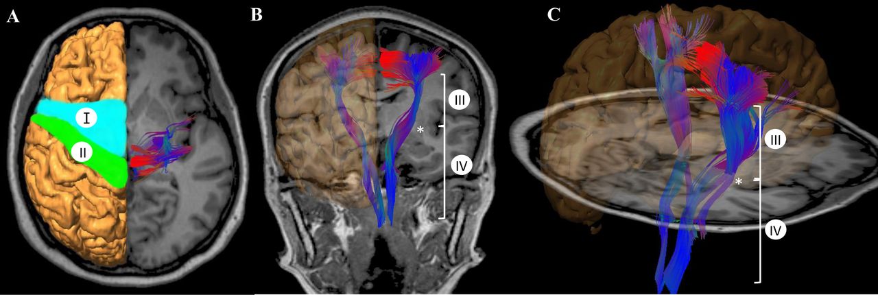
According to previous studies and clinical practice, four motor-related area subtypes were identified based on nidus location: (I) nidus involving PMA and/or SMAs; (II) nidus involving the precentral gyrus; (III) nidus involving the CST and superior to the posterior limb of the internal capsule; (IV) nidus that invaded the CST at or inferior to the level of posterior limb of the internal capsule.7 If more than one motor-related area was invaded, the nidus was sequentially classified as the highest number of the subtypes (figure 1). In addition, the deep perforating artery supply was taken into consideration. We divided type IV into type IVa (nidus without AChA feeding) and type IVb (nidus with AChA feeding). The nidus location was identified using TOF-MRA and DTI tractography images. Nidus was defined as involving the CST if the shortest distance from a nidus to the CST was less than 5 mm based on DTI tractography.

Classification of motor-related eloquent areas on different planes. (A) On axial plane; (B) on coronal plane; (C) on sagittal plane. Type I, nidus involving premotor area and/or supplementary motor areas; type II, nidus involving the precentral gyrus; type III, nidus involving the corticospinal tract (CST) and superior to the posterior limb of the internal capsule; type IV, nidus that invaded the CST at or inferior to the level of posterior limb of the internal capsule. Asterisk refers to the level of internal capsule.
Neuroimaging
Sagittal T1-weighted anatomical MR images, time-of-flight (TOF) MRA and DT images were obtained using a 3.0 T MR scanner (SIEMENS Trio) as previously reported.11 The acquired image data were analysed on the iPlan cranial 3.0 workstation (Brainlab). Image co-registration and fusion were performed using an automatic rigid registration. Two regions of interest delineated in the precentral gyrus (seed) and pons (target) were used to track the CST.12
Surgery
Surgical removal of AVMs was performed by an experienced physician (YC). A neuronavigation system was used to help preserve white matter tracts. Intraoperative electrophysiology monitoring and motor area mapping were used to identify the motor cortex and CST. Intraoperative indocyanine fluorescence angiography and ultrasonography were used to discern the feeding arteries and nidus margin of AVMs. Digital subtraction angiography (DSA) was performed after surgery to validate radical obliteration.
Muscle strength evaluation
Muscle strength assessments (Medical Research Council Scores 0–5) were performed 1 day preoperatively, 1 week postoperatively (short-term) and up to 6 months postoperatively (long-term). Follow-up information from the period during the recovery of motor function was obtained by telephonic interviews. The recovery time of the motor deficits was recorded. The standard of recovery was defined as patients recovering to the level of being able to carry out all usual activities despite some symptoms (modified Rankin Scale (mRS) ≤1). Long-term good outcomes were defined as mRS 0–2, and poor outcomes were defined as mRS >2 at 6 months. The surgical complications in this study included intracranial infection, rehaemorrhage and cerebral infarction.
Variables
The nidus size, eloquence, S-M grade, and presence of deep venous drainage and deep perforating arterial feeders were determined through preoperative angiograms.13 Deep perforating arteries included the medial lenticulostriates, lateral lenticulostriate arteries, anterior and posterior choroidal arteries, thalamoperforators and brainstem perforators.14 Haemorrhagic presentation was defined as radiographic evidence of haemorrhage on CT or MRI. Diffuseness was determined from preoperative angiograms and using TOF to identify intervening brain parenchyma within the nidus.
Statistical analyses
Statistical analyses were performed with SPSS V.20.0.0. Categorical variables are summarised as frequency counts and percentages and were compared using the χ2 test or Fisher’s exact test. Continuous variables are summarised as means±SD and were compared using an independent samples t-test. Postoperative short-term and long-term mRS scores were analysed to identify the surgical outcomes. The association between the variables and surgery-related motor deficits (MDs) was analysed using univariate and multivariate analyses. Variables with p value <0.1 in the univariate analysis were then used in the multivariate analysis model. For the recovery time of motor deficits, Kaplan-Meier survival analysis was applied to illustrate the time-to-recover curves. The log-rank test was used to compare differences among motor-related area subtypes. Statistical tests were considered significant at p value <0.05. ORs and HRs are presented with 95% CIs.
Results
Clinical characteristics
A total of 125 patients with M-AVM were included in this study (table 1). There were 69 (55.2%) male patients and 56 (44.8%) female patients, with a mean age of 26.3±13.4 years. There were 12 (9.6%), 44 (35.2%), 57 (45.2%) and 12 (9.6%) patients scored S-M grade I, II, III and IV, respectively. Thirty-three (26.4%) patients had preoperative haemorrhages. The mean nidus diameter was 36.8 mm (5.0–93.6 mm). Forty patients (32.0%) had a diffuse nidus, and 85 patients (68%) had a compact nidus. The number of patients with nidus involving I, II, III and IV were 50 (40.0%), 25 (20.0%), 34 (27.2%) and 16 (12.8%), respectively.
Demographic and angioarchitectural characteristics of patients with M-AVMs
There were no statistically significant differences in terms of the patient age (p=0.270) and sex (p=0.403) among the different subtypes. For the AVM characteristics, deep perforating arteries were more common in type III (41.2%) and type IV (81.2%) AVM cases (p<0.001). Deep venous drainage was more common in type IV AVM cases (50%, p<0.001). There were no differences in nidus diffuseness, size (p=0.334), haemorrhage (p=0.148) and post-surgical complications (p=0.350) among these four subtype groups.
Preoperative seizure
In our cohort, the incidence of preoperative epilepsy differed among the four subtypes. There were 31 (62.0%) in type I patients, 12 (48.0%) in type II patients, 12 (35.3%) in type III patients and 7 (43.8%) in type IV patients with an epileptic history before surgery. For the seizure types, patients in the type I group had a higher incidence (56%) of generalised tonic–clonic seizures than patients in the other groups (online supplemental table S1). Table 2 summarizes the preopeartive antiepileptic drug use and the indications for surgery.
Supplemental material
Preoperative antiepileptic drug use and the indications for surgery
Risk factors for MDs
Radical obliteration was achieved in all patients according to postoperative DSA. Sixty-one (48.8%) patients suffered from short-term MDs 1 week after surgery. According to the univariate analysis, significant differences in motor-related area subtypes (p<0.001), diffuseness (p=0.004), size (p=0.010), deep perforating artery supply (p=0.032) and S-M grade (p<0.001) were found between the patient cohorts with and without postoperative short-term MDs. No significant difference was found in the other factors (online supplemental table S2). According to the multivariate analysis, motor-related area subtypes (p=0.003) and diffuseness (p=0.042) were significantly associated with an increased risk of short-term MDs. No significant difference was found in the other factors (table 3).
Multivariate logistic regression analysis of predictors of postoperative motor deficits (MDs)
At the follow-up after 6 months, 45 (36.0%) patients suffered from different levels of long-term MDs. Only 10 (7.8%) patients had a mRS >2 as a poor outcome. According to the univariate analysis, significant differences in motor-related area subtypes (p=0.002), diffuseness (p=0.008), S-M grade (p=0.027), deep perforating artery supply (p=0.038) and deep venous (DV) drainage (p=0.044) were found between the patient cohorts with and without postoperative MDs. No significant difference was found in the other factors (table 4). According to the multivariate analysis, motor-related area subtypes (p=0.004) and diffuse nidus (p=0.014) were significantly associated with an increased risk of motor deficits. We compared the predictive accuracy of the motor-related area subtypes and S-M grade by constructing receiver operating characteristic (ROC) curves. A higher area under ROC curve (0.70 vs 0.63) of the new classification than SM grade was observed (online supplemental figure S1). No significant difference was found in the other factors (table 3).
Supplemental material
Univariate analysis of predictors of postoperative long-term motor deficits (MDs)
Proportion of MDs
Different proportions of short-term (p<0.001) and long-term (p=0.005) MDs were observed among the four subtypes. The post-surgical short-term MDs were significantly more frequent in type III patients than in type I (p<0.001) or type II (p=0.047) patients. The post-surgical short-term MDs were significantly less frequent in type I patients than in type II (p=0.009), type III or type IV (p=0.002) patients (online supplemental table S2). For long-term MDs, post-surgical MDs were significantly less frequent in type I patients than in type II (p=0.009), type III (p<0.001) or type IV (p=0.004) patients. Although the type IV patients did not suffer from the highest proportion of postoperative MDs, they acquired the highest proportion (four patients, 25.0%) of long-term poor outcomes (mRS >2) compared with type I (two patients, 4.0%), type II (0 patients, 0%) and type III (four patients, 11.8%) patients. The difference between type IV and type I (p=0.041) patients and type IV and type II (p=0.018) patients was significant (figure 2). In addition, type IVb patients showed a significantly higher incidence of post-surgical MDs than type IVa (p=0.041) patients (table 5).

Poor surgical outcomes of arteriovenous malformations located in motor-related areas involving different motor-related area subtypes. The x-axis indicates the motor-related area subtypes. The y-axis indicates the percentage of good and poor surgical outcomes (*p<0.05).
Comparison of surgical outcomes of patients with type IV M-AVMs
Muscle strength recovery time
The muscle strength recovery time differed in different subtypes (figure 3). The mean recovery time was 0.96±2.0 months in type I, 1.72±2.3 months in type II, 3.2±2.6 months in type III and 3.6±2.6 months in type IV patients. A Mann-Whitney U test revealed that compared with that of type I or II patients, the muscle strength of type III or IV patients required more days to recover (type I vs III, p<0.001; type II vs III, p=0.029; type I vs IV, p<0.001; type II vs IV, p=0.018).

Recovery time of motor deficits in arteriovenous malformations (AVMs) located in motor-related areas involving different motor-related area subtypes. (A) Kaplan-Meier survival plots for AVMs according to months of motor deficit recovery. The x-axis indicates completed months of follow-up. (B) Bar graph showing the average recovery time of different motor-related area subtypes (*p<0.01; ***p<0.001).
Postoperative seizure control
With regular antiepileptic drugs, of the 62 patients who had a preoperative seizure history, 90.3% of these patients (56/62) controlled their seizures well and reached Engel class I after tumour resection. Three patients (4.8%) were classified as Engel class II. Two patients (3.2%) were classified as Engel class III. Moreover, one patient (1.6%) was classified as Engel class IV. There were no significant differences among these four subtypes.
Discussion
The present study confirms our hypothesis that AVM motor eloquence subtype does have an influence on outcome following resection. We found that type I AVMs may strengthen the recommendation for surgery considering the high incidence of preoperative seizure, the relatively low rate of post-surgical MDs and the satisfactory post-surgical seizure control. When the nidus was involving CST at or below the level of posterior limb of the internal capsule, especially fed by the AChA, the microsurgical treatment decision should be prudently appraised in light of the surgical risk and natural history of AVMs.
Postoperative functional MDs
According to present data, post-surgical motor status was different among the subtypes. A high incidence of poor outcomes were observed in patients with type IV AVMs. We attribute it to two main reasons. First, according to our previous data, the CST level involved may be associated with surgical outcomes. CST rupture below the corona radiata seemed to be correlated with poor long-term outcomes. The sample size in that study was small (only 1).15 Our present data validated the conclusion with a larger sample size. Second, a higher incidence of deep perforating arteries and DV drainage was observed in type IV AVMs than in the other types. According to the literature, the DV drainage and perforating artery supply are friable, resist bipolar coagulation and have the dangerous propensity to retract. Their deep location can limit visualisation and overall operative manoeuvrability, and adds a unique set of dangers to AVM resection.16 In our study, there were 14 (41.2%) type III and 13 (81.2%) type IV patients with nidus existing in deep perforating arteries, while there were 3 (8.8%) and 8 (50.0%) patients with existing DV drainage. Accordingly, in our opinion, the poor outcomes in type III patients were more associated with damage to the adjacent eloquent areas, while type IV patients were associated with both damage to adjacent eloquent areas and angioarchitectural characteristics.
In this study, 22% (11/50) of type I patients suffered from short-term motor strength deficits. This could be due to postoperative supplementary motor area syndrome.17 The PMA and SMA contribute significantly to the control of hand movements required for the manipulation of objects.18 In 16% (8/60) of type I patients, there was no recovery of MDs to normal 6 months after surgery. The proportion was lowest among the four subtypes but still higher than that of glioma resection in this area. In previous studies, infarction or resection of the PMA or SMA in gliomas often leads to weakness of muscles or akinesia, which can resolve spontaneously and completely within days to months.19–21We speculated that the delayed recovery was associated with resection of the cingulate cortex and its deep regions or interruption of the CST.16 Different from cerebral infarctions or gliomas, in surgery for AVMs, rough nidus margins and intermixed adjacent brain tissue force the surgeon to damage the functional fibres when resecting the nidus.
Subtypes of type IV
In our previous study, we described that surgical treatment of AVMs supplied by the AChA, especially the cisternal segment, can cause a high incidence of MDs.6 Therefore, we divided type IV into type IVa (nidus without AChA feeding) and type IVb (nidus with AChA feeding). Consistent with our speculation, a significant difference in long-term MDs was observed between the two groups. In addition to the anatomy location, these data indicated that the feeding arteries have a great effect on the surgical outcomes of M-AVMs. The AChA nourishes many important anatomical structures such as the internal capsule, the lateral geniculate body and the thalamus.12 13 During the surgical treatment of M-AVMs, the feeding arteries must be ligated to facilitate the resection.22 Brain AVMs supplied by cisternal segment of AChA was an independent risk factor for postoperative MDs because the perforating branches of the cisternal segment of the AChA do not receive any significant collateral supply.6 In this study, patients with a nidus located in a type IVb area was mostly fed by the cisternal part of the AChA, so the effect of AChA feeding is mainly affecting MDs of type IV patients.
Recovery time
According to present data, the recovery time after surgery differed among different subtypes. Patients with type I AVMs needed the shortest recovery thanks to the mild motor deficits. In contrast, patients with type III or type IV AVMs needed the longest recovery time resulting from the relatively severe MDs. It was worth mentioning that patients with AVMs involving motor fibres (types III and IV) needed longer recovery times than those with AVMs involving the motor cortex (type II), although they were both primary eloquent motor areas. We attributed it to two reasons. The first one is the different recovery power between the motor cortex and subcortical CST. According to the literature, distinct to the cortex, subcortical pathways are difficult to compensate for, especially pathways responsible for a single function. Therefore, the rupture of the CST may need a longer time to reorganise.23 Second, we speculate that this difference is also a result of the larger potential of cortical reorganisation in patients with type II AVMs. There is a consensus that AVMs develop in early life.24 According to previous studies, the brain cortex has great ability to reorganise in the setting of a chronic disease, with displacement to homologous regions on the contralateral side or adjacent regions on the ipsilateral side.25 26 The reorganisation may facilitate motor recovery after the surgery-related injury of the brain cortex.
Epileptic status
A high incidence (62%) of preoperative seizures was observed in type I AVMs, which is higher than that of seizures due to frontal AVMs reported in the literature (39%). In a previous study, Hamasaki et al demonstrated the higher incidence of epilepsy in meningiomas located on the PMA.27 In addition, Wang et al demonstrated that increased seizure risks were identified for low-grade gliomas that involved the PMA using voxel-based lesion-symptom mapping.28 Literature about the epileptic status of patients with AVMs located in the PMA or SMA were limited to case reports. Our study showed the high incidence of epilepsy in these areas. After surgery, satisfactory seizure control was achieved. In our series, 88% of patients with type I AVMs who underwent surgery were seizure free or Engel class I at the 6-month follow-up.
Other factors related to post-surgical MD
According to present data, nidus diffuseness was independently associated with short-term and long-term MDs. This outcome was in concordance with previous studies.14 29 30 Compact M-AVMs have distinct borders so that clear dissection can be achieved between brain tissue and nidus, whereas a diffuse nidus can force the surgeon to dissect too close to the nidus, resulting in unexpected bleeding, or resect interspersed brain, which might injure the eloquent motor cortex or CST.16
Study limitations
Our study had several limitations. First, this study was conducted in a single centre. Second, due to its retrospective nature, it may be difficult to avoid information bias, selection bias and confounding factors. Third, although the sample size in this study may be one of the largest regarding the study of M-AVMs, to the best of our knowledge, the sample size was still not large enough because there were only 16 patients in the type IV group. Meanwhile, the limited number of outcomes may result in overfitting of the model when the statistical model contains more parameters than can be justified by the data. Thus, further validation with more patients and a prospective approach should be performed.
Conclusions
The motor-related area classification system for M-AVMs provides an observational tool in patients with M-AVMs. Based on our results and the literature, we propose some suggestions for patient selection in surgically treating this challenging entity. Considering the high incidence of preoperative seizure (especially the generalised tonic–clonic subtype), the satisfying post-surgical seizure control and the relatively low rate of post-surgical MD, the recommendation for surgery of type I AVMs may be strengthened. Great caution should be taken in the resection of type II and III AVMs to avoid postoperative MDs, especially type III. A longer recovery time was needed for type III AVMs than for type II AVMs. Moreover, the AChA feeding should also be taken into consideration when classifying M-AVMs. For the type IV AVMs, especially type IVb, because of the high proportion of poor post-surgical outcomes, the microsurgical treatment decision should be prudently appraised in light of the surgical risk and natural history of AVMs.31 In our opinion, conservative treatment may be recommended for type IVb patients without a history of bleeding or intractable seizure. If the surgery were chosen for the aforementioned reasons, radiosurgery, embolisation, or a combination of microsurgery and embolisation can be recommended, whereas the benefits of these treatment modalities require validation in further research.
Data availability statement
Data are available on reasonable request. Deidentified participant data are available on reasonable request.
Ethics statements
Patient consent for publication
Ethics approval
This study complied with the Declaration of Helsinki, adhered to the Good Clinical Practice guideline and was approved by the Ethics Committee of the Beijing Tiantan Hospital Affiliated with Capital Medical University (KY2016-031-01). Written informed consent was obtained from all participants or their legally authorised representative.
References
Supplementary material
Supplementary Data
This web only file has been produced by the BMJ Publishing Group from an electronic file supplied by the author(s) and has not been edited for content.
Footnotes
Contributors Conception and design: YC, YJ, TJ. Acquisition of data: YJ, JW, YW, WF. Analysis and interpretation of data: YJ, YW, HL, YC. Drafting the article: YJ. Critically revising the article: YC, YJ, TJ. Reviewed submitted version of manuscript: all authors. Approved the final version of the manuscript on behalf of all authors: YC, TJ. Statistical analysis: YJ, YW, HL. Administrative/technical/material support: YC, TJ, SW, JZZ. Study supervision: YC, TJ, JZZ.
Funding This study was supported by the National Natural Science Foundation of China (Grant No. 81901175, Principal Investigator: YJ), the “National key research and development program of China during the 13th Five-Year Plan Period” (Grant No. 2016YFC1301803, Principal Investigator: YC; and Grant No. 2016YFC1301801, Principal Investigator: SW).
Competing interests None declared.
Provenance and peer review Not commissioned; externally peer reviewed.